Narciso F. Atienza, Jr. MD, MBA, FPCS, FPAO
EYE PHYSICIAN AND SURGEON
FELLOW, PHILIPPINE COLLEGE OF SURGEONS
FELLOW, PHILIPPINE ACADEMY OF OPHTHALMOLOGY
DIPLOMATE, PHILIPPINE BOARD OF OPHTHALMOLOGY
SUBSPECIALTY IN DISEASES AND SURGERY OF THE RETINA, VITREOUS, AND MACULA and OCULAR ONCOLOGY
FELLOW, PHILIPPINE COLLEGE OF SURGEONS
FELLOW, PHILIPPINE ACADEMY OF OPHTHALMOLOGY
DIPLOMATE, PHILIPPINE BOARD OF OPHTHALMOLOGY
SUBSPECIALTY IN DISEASES AND SURGERY OF THE RETINA, VITREOUS, AND MACULA and OCULAR ONCOLOGY

What is a retinal artery occlusion?
Retinal artery occlusion is a blockage in one of the small arteries that carry blood to the retina. The retina is a layer of tissue in the back of the eye that is able to sense light.
What causes a retinal artery occlusion?
Retinal arteries may become blocked by a blood clot or fat deposits that get stuck in the arteries. These blockages are more likely if there is hardening of the arteries (atherosclerosis) in the eye.
Clots may travel from other parts of the body and block an artery in the retina. The most common sources of clots are the carotid artery in the neck and the heart.
Most clots occur in people with conditions such as:
Carotid artery disease, a condition in which the two large blood vessels in the neck become narrowed or blocked
Diabetes
Heart rhythm problem (atrial fibrillation)
Heart valve problem
High levels of fat in the blood (hyperlipidemia)
High blood pressure
Intravenous drug abuse
Temporal arteritis (damage to arteries due to an immune response)
If a branch of the retinal artery is blocked, part of the retina will not receive enough blood and oxygen. If this happens, you may lose part of your vision.
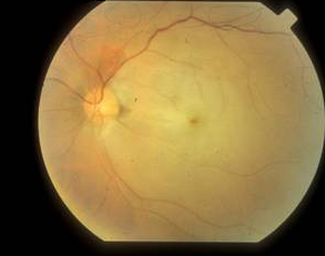
Eye with Branch Artery Occlusion
Eye with Central Artery Occlusion
What are the symptoms of an artery occlusion?
Sudden painless blurring or loss of vision may occur in:
All of one eye (central retinal artery occlusion or CRAO)
Part of one eye (branch retinal artery occlusion or BRAO)
The retinal artery occlusion may last for only a few seconds or minutes, or it may be permanent.
If the blood clot moves to another part of the brain, symptoms of a stroke may develop.
What tests or procedures are needed to be done?
Tests to evaluate the retina may include:
Examination of the retina after dilating the pupil
Fluorescein angiography
Intraocular pressure
Pupil reflex response
Refraction
Retinal photography
Slit lamp examination
Testing of side vision (visual field examination)
Visual acuity
General tests should include:
Blood pressure
Blood tests, including cholesterol and triglyceride levels and the erythrocyte sedimentation rate
Physical examination
Tests to identify the source of a clot from another part of the body:
Echocardiogram
Electrocardiogram
Heart monitor for abnormal heart rhythm
Duplex Doppler ultrasound of the carotid arteries
What is the treatment for an artery occlusion?
There is no proven treatment for vision loss that involves the whole eye, unless it is caused by another illness that can be treated.
Several treatments may be tried. These treatments must be given within 2 - 4 hours after symptoms begin to be helpful. However, the benefit of these treatments has never been proven, and they are rarely used.
Breathing in (inhaling) a carbon dioxide-oxygen mixture. This treatment causes the arteries of the retina to widen (dilate).
Massage of the eye
The clot-busting drug, tissue plasminogen activator (tPA)
The health care provider should look for the cause of the blockage. Blockages may be signs of a life-threatening medical problem.
Retinal artery occlusion is a blockage in one of the small arteries that carry blood to the retina. The retina is a layer of tissue in the back of the eye that is able to sense light.
What causes a retinal artery occlusion?
Retinal arteries may become blocked by a blood clot or fat deposits that get stuck in the arteries. These blockages are more likely if there is hardening of the arteries (atherosclerosis) in the eye.
Clots may travel from other parts of the body and block an artery in the retina. The most common sources of clots are the carotid artery in the neck and the heart.
Most clots occur in people with conditions such as:
Carotid artery disease, a condition in which the two large blood vessels in the neck become narrowed or blocked
Diabetes
Heart rhythm problem (atrial fibrillation)
Heart valve problem
High levels of fat in the blood (hyperlipidemia)
High blood pressure
Intravenous drug abuse
Temporal arteritis (damage to arteries due to an immune response)
If a branch of the retinal artery is blocked, part of the retina will not receive enough blood and oxygen. If this happens, you may lose part of your vision.
Eye with Branch Artery Occlusion
Eye with Central Artery Occlusion
What are the symptoms of an artery occlusion?
Sudden painless blurring or loss of vision may occur in:
All of one eye (central retinal artery occlusion or CRAO)
Part of one eye (branch retinal artery occlusion or BRAO)
The retinal artery occlusion may last for only a few seconds or minutes, or it may be permanent.
If the blood clot moves to another part of the brain, symptoms of a stroke may develop.
What tests or procedures are needed to be done?
Tests to evaluate the retina may include:
Examination of the retina after dilating the pupil
Fluorescein angiography
Intraocular pressure
Pupil reflex response
Refraction
Retinal photography
Slit lamp examination
Testing of side vision (visual field examination)
Visual acuity
General tests should include:
Blood pressure
Blood tests, including cholesterol and triglyceride levels and the erythrocyte sedimentation rate
Physical examination
Tests to identify the source of a clot from another part of the body:
Echocardiogram
Electrocardiogram
Heart monitor for abnormal heart rhythm
Duplex Doppler ultrasound of the carotid arteries
What is the treatment for an artery occlusion?
There is no proven treatment for vision loss that involves the whole eye, unless it is caused by another illness that can be treated.
Several treatments may be tried. These treatments must be given within 2 - 4 hours after symptoms begin to be helpful. However, the benefit of these treatments has never been proven, and they are rarely used.
Breathing in (inhaling) a carbon dioxide-oxygen mixture. This treatment causes the arteries of the retina to widen (dilate).
Massage of the eye
The clot-busting drug, tissue plasminogen activator (tPA)
The health care provider should look for the cause of the blockage. Blockages may be signs of a life-threatening medical problem.