Narciso F. Atienza, Jr. MD, MBA, FPCS, FPAO
EYE PHYSICIAN AND SURGEON
FELLOW, PHILIPPINE COLLEGE OF SURGEONS
FELLOW, PHILIPPINE ACADEMY OF OPHTHALMOLOGY
DIPLOMATE, PHILIPPINE BOARD OF OPHTHALMOLOGY
SUBSPECIALTY IN DISEASES AND SURGERY OF THE RETINA, VITREOUS, AND MACULA and OCULAR ONCOLOGY
FELLOW, PHILIPPINE COLLEGE OF SURGEONS
FELLOW, PHILIPPINE ACADEMY OF OPHTHALMOLOGY
DIPLOMATE, PHILIPPINE BOARD OF OPHTHALMOLOGY
SUBSPECIALTY IN DISEASES AND SURGERY OF THE RETINA, VITREOUS, AND MACULA and OCULAR ONCOLOGY

What is a retinal vein occlusion?
Retinal vein occlusion occurs when the circulation of a retinal vein becomes obstructed by an adjacent blood vessel, or by a small blood clot inside the vein itself. The retina is the light sensing tissue at the back of the eye. The veins drain blood out of the retina and return it to the heart.
Blockage or occlusion in the vein prevents adequate blood flow in the affected area (Ischemia). The walls of the vein leak blood and excess fluid into the retina (Swelling).
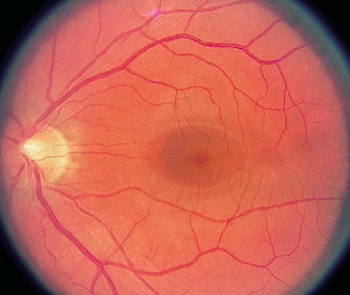
Eye with a Central Retinal Vein Occlusion
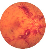
Eye with a branch retinal vein occlusion
Swelling and ischemia (lack of oxygen) of the retina as well as glaucoma are fairly common complications.
What are its’ symptoms?
The visual symptoms can vary in severity from one person to the next, and are dependent on whether the central retinal vein or a branch retinal vein is involved. Patients who experience a branch vein occlusion often notice a gradual improvement in their vision as the hemorrhage resolves. Recovery from a central vein occlusion is much less likely since it affects the macula. Symptoms may include: 1) Sudden blurring of vision, 2) Decreased vision in a certain part of the vision (branch vein occlusion, 3) Total decreased in vision (central vein occlusion)
Does this problem occur very often?
This problem appears equally in males and females and is more common after the age of 60. These disorders are similar to those for vascular occlusive disease elsewhere in the body such as stroke and coronary artery disease. Specifically, aging, high blood pressure, diabetes, and smoking are all risk factors. When these problems occur in young patients, a history of oral contraceptive use, or history of drug abuse, or a history of blood problems can be elicited in some patients though majority will be negative on any of the aforementioned conditions.
How is it detected and diagnosed?
Vein occlusion is diagnosed by examining the retina with an ophthalmoscope. Fluorescein angiography may be performed in some cases to study the circulation of the retina and to determine the extent of macular edema or swelling. This is usually done when the hemorrhages have cleared up, as doing angiography on an acutely congested eye with occlusion will not give any information. An O.C.T. (Optical coherence topography) may give information on the involvement of the macula.
Young patients, basing on history should be investigated for blood problems, or any medication intake, like oral contraceptive pills. A consult with your family doctor or internist may be necessary to find out the cause and risks for this condition.
Treatment.
There is no cure for a vein occlusion. Following a vein occlusion, the primary concern is to treat the secondary complications. If areas of the retina are oxygen-deprived as seen with an angiogram, close observation is usually recommended to watch out for the growth of new vessels. Angiography can also show areas of swelling and leakage. LASER is recommended when new vessels are present and are at risk of bleeding or developing neo-vascular glaucoma, or if there is significant swelling on the macula.
Other treatment modalities have been investigated recently like the injection of medications (steroids, anti-VEGF) inside the eye, and vitrectomy surgery to try to decompress the blocked vein or to create another way for the stagnant blood to flow thru. These options have been shown to be successful in alleviating vein occlusion in a small series of patients.
None of the treatment options will bring back your full vision. It is hoped that these options will limit or even halt the progressive visual loss associated with retinal vein occlusions.
Your retina surgeon will advise you on the best course for your condition, whether he will request for a fluorescein angiogram or an OCT, do observation with close follow-up, laser, injection of medications or even vitreous surgery.
Cataract surgery alone will not cause any remarkable improvement if done. It may even cause worsening of vision over time.
Why are regular medical eye examinations important for everyone?
Eye diseases can strike at any age. Many eye diseases do not cause symptoms until the disease has done damage. Since most blindness is preventable if diagnosed and treated early, regular medical examinations by an ophthalmologist are very important.
Retinal vein occlusion occurs when the circulation of a retinal vein becomes obstructed by an adjacent blood vessel, or by a small blood clot inside the vein itself. The retina is the light sensing tissue at the back of the eye. The veins drain blood out of the retina and return it to the heart.
Blockage or occlusion in the vein prevents adequate blood flow in the affected area (Ischemia). The walls of the vein leak blood and excess fluid into the retina (Swelling).
Eye with a Central Retinal Vein Occlusion
Eye with a branch retinal vein occlusion
Swelling and ischemia (lack of oxygen) of the retina as well as glaucoma are fairly common complications.
What are its’ symptoms?
The visual symptoms can vary in severity from one person to the next, and are dependent on whether the central retinal vein or a branch retinal vein is involved. Patients who experience a branch vein occlusion often notice a gradual improvement in their vision as the hemorrhage resolves. Recovery from a central vein occlusion is much less likely since it affects the macula. Symptoms may include: 1) Sudden blurring of vision, 2) Decreased vision in a certain part of the vision (branch vein occlusion, 3) Total decreased in vision (central vein occlusion)
Does this problem occur very often?
This problem appears equally in males and females and is more common after the age of 60. These disorders are similar to those for vascular occlusive disease elsewhere in the body such as stroke and coronary artery disease. Specifically, aging, high blood pressure, diabetes, and smoking are all risk factors. When these problems occur in young patients, a history of oral contraceptive use, or history of drug abuse, or a history of blood problems can be elicited in some patients though majority will be negative on any of the aforementioned conditions.
How is it detected and diagnosed?
Vein occlusion is diagnosed by examining the retina with an ophthalmoscope. Fluorescein angiography may be performed in some cases to study the circulation of the retina and to determine the extent of macular edema or swelling. This is usually done when the hemorrhages have cleared up, as doing angiography on an acutely congested eye with occlusion will not give any information. An O.C.T. (Optical coherence topography) may give information on the involvement of the macula.
Young patients, basing on history should be investigated for blood problems, or any medication intake, like oral contraceptive pills. A consult with your family doctor or internist may be necessary to find out the cause and risks for this condition.
Treatment.
There is no cure for a vein occlusion. Following a vein occlusion, the primary concern is to treat the secondary complications. If areas of the retina are oxygen-deprived as seen with an angiogram, close observation is usually recommended to watch out for the growth of new vessels. Angiography can also show areas of swelling and leakage. LASER is recommended when new vessels are present and are at risk of bleeding or developing neo-vascular glaucoma, or if there is significant swelling on the macula.
Other treatment modalities have been investigated recently like the injection of medications (steroids, anti-VEGF) inside the eye, and vitrectomy surgery to try to decompress the blocked vein or to create another way for the stagnant blood to flow thru. These options have been shown to be successful in alleviating vein occlusion in a small series of patients.
None of the treatment options will bring back your full vision. It is hoped that these options will limit or even halt the progressive visual loss associated with retinal vein occlusions.
Your retina surgeon will advise you on the best course for your condition, whether he will request for a fluorescein angiogram or an OCT, do observation with close follow-up, laser, injection of medications or even vitreous surgery.
Cataract surgery alone will not cause any remarkable improvement if done. It may even cause worsening of vision over time.
Why are regular medical eye examinations important for everyone?
Eye diseases can strike at any age. Many eye diseases do not cause symptoms until the disease has done damage. Since most blindness is preventable if diagnosed and treated early, regular medical examinations by an ophthalmologist are very important.